
Pleural Diseases
Einstein Health Glossary
ICD 10 - J90-J94

ICD 10 - J90-J94
The pleura is a thin layer of tissue that lines the lungs (visceral pleura) and the inner wall of the chest (parietal pleura). In healthy individuals, there is a constant flow of fluid between these two thin layers. However, pleural diseases can lead to an abnormal accumulation of fluid between the pleurae, known as pleural effusion. The most common causes of pleural effusion include: tuberculosis, cancer, and pneumonia.
Pleural effusion is the excessive accumulation of fluid in the pleural space (between the lung and the ribs).
VATS – Video-Assisted Thoracic Surgery can be performed to treat this condition in the following ways:
These procedures allow the lung to return to its normal position and help reduce respiratory symptoms.
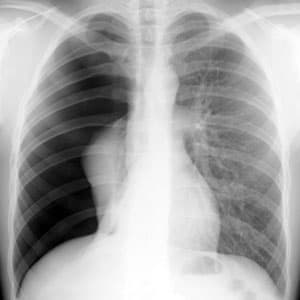
Pneumothorax occurs when air escapes into or enters the pleural space (between the lung and the ribs), causing a partial or complete collapse of the lung.
VATS – Video-Assisted Thoracic Surgery is performed to treat and prevent the recurrence of this condition, especially when a previous treatment with pleural drainage is insufficient.
During the procedure: